OVERVIEW
X-ray, also called general or diagnostic radiography, is an extremely valuable and common tool in the diagnosis of internal injuries and other medical conditions.
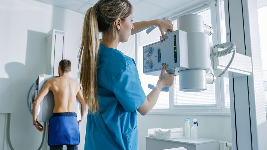
X-ray exams begin by sending beams of energy through the body. Bones and other dense materials (like a foreign object inside the body) will absorb more of this energy than organs and soft tissues – creating clearer images of concentrated energy. The X-ray camera detects the energy and produces pictures of the internal structures. The bones appear white or gray on the x-ray image, while soft tissues will appear darker.
Fluoroscopy is an X-ray technique that captures moving images of internal structures on a video monitor. Radiologists use it to view organs in motion and assess their function with contrast material. It also guides needle placement during myelography and joint injections.
What to Expect during an X-Ray Exam
Patient Testimonials
Brian
“My XRAY tech ‘Brian’ was very personable and professional…”
Shyla
“Receptionist Shyla was particularly helpful. I was in for a kidney stone x-ray… Shyla helped me figure out who the doc was… and got a verbal order.”
Sierra, Allyson
“Foot x-rays – Rad techs Sierra and Allyson were especially nice.”
Pam
“Had a patient call wanting to tell you what an amazing job Pam did… Said the x-ray should probably really be a STAT.”
Frequently Asked Questions
X-ray, also called general or diagnostic radiography, is a common imaging tool that uses energy beams to create pictures of your internal structures. Bones and dense materials appear white or gray, while soft tissues appear darker on the images.
Fluoroscopy is a specialized X-ray technique that captures real-time moving images of your internal structures, like a movie. It allows radiologists to view organs in motion and assess their function, making it ideal for procedures that require live guidance, such as joint injections and myelography.
Most general radiography exams take between 10 to 30 minutes, depending on the type of exam or procedure being performed.
Preparation is typically minimal. You may be asked to change into a gown if your clothing has zippers or buttons that could interfere with the imaging.
General radiography exams usually take between 10 to 30 minutes, depending on the type of exam/procedure. You may be asked to change into a gown if your clothing has zippers or buttons.
Usually, there are no restrictions following a typical X-ray exam. However, if you receive a fluoroscopy exam, such as a myelogram of the spinal cord, you may receive some post-exam instructions.
Like all imaging exams, a radiologist will review your results and compare your exam to any of your previous studies that are applicable. A report will be then be created and sent to your physician.
Most of the time, your physician’s office will work directly with Inland Imaging to schedule an appointment for you. If you have been asked to make your own appointment, contact Inland Imaging for scheduling information at (509) 455-4455 or toll-free at 1.800.826.2944. Before calling, be sure your physician’s order and insurance information (including pre-authorization, if needed) are readily available. Women who may possibly be pregnant should notify the technologist immediately before receiving the exam.